TIME

الاثنين، مايو 11

Episode 6

Common injuries of the cervical spine
.
.
Penetrating injury such as a gunshot wound that may have traversed the spine
.
Hangman's Fracture - caused by simultaneous extension and distraction as in being “clothes lined” at high speed, or during a MVA in which the head is forced against the car roof creating compression followed by an extension vector
.
.
Odontoid Fractures - are classified as type I, type II, or type III fracture 
..
Type I odontoid fractures involve only the tip of the dens and are mostly considered stable
.
Type II fractures are the most common type involving the dens and body of C-2 vertebra. Following reduction the patient placed in a halo and/or surgical fusion may be necessary 
.
Type III odontoid fractures have deep nonunion into the body of C2 vertebra 
.
.
Jefferson Fracture - is a type of burst fracture of C-1 vertebra. It can occur from an acutely severe axial force causing compression. If the force is great as in diving injuries, the vertebral arch or body of adjacent vertebra literally burst. In the absence of bone fragments penetrating the spine survival without neurological deficit occurs in about 50% of patients, especially if the lateral masses are forced laterally away from the spinal cord
.
.
Rotational Injury - Involves complex forces caused by injury onto the head or neck. When rotational injuries involve hyperextension, complex fractures to the interarticularis and pedicle(s) may occur. A "jumped facet(s) phenomenon man occur either unilateral or bilaterally. In some cases the subluxation may be unstable such as when the facets are jumped but not locked
.
.
Extension teardrop fractureCaused by an avulsion of the anterior part of a vertebral body during hyperextension
.
.
Clay-shoveler’s fractureA fracture of the spinous process of C6, C7, or T1. It is due to hyperflexion injury and may involve one or more spinous process
.
.
Unilateral locked facetcaused by flexion and rotation injury. The intervertebral foramina is visible above but no below on the true lateral view
.
.
Bilateral locked facetsalso caused by hyperflexion injury. The upper cervical spine is anteriorly displaced over the lower spine. A clue to this injury is widening of the spinous processes at the site of injury. This results in a complete luxation (dislocation) of the spine
.
.
Muscle spasm: loss of normal lordotic curve (lateral view), or torticollis on AP view
.
.
Truescene is your right choice


الأحد، مايو 10

Episode 5

Plain Film Radiographic Imaging of the Traumatic C-spine


Trauma imaging of the cervical spine begins with a high suspicion for vertebral or spinal cord injury. Standard radiographic views are obtained with the patient supine, fully supported on a spine board, with neck collar and immobilization apparatus in place. The standard trauma film series at most institutions are: a horizontal beam lateral and Swimmer's view (if necessary), anterior-posterior (AP) view, and open mouth odontoid view
.
Diagnostic imaging technologists should be aware of the indications for special views of the cervical spine such as: inability to adequately visualize all vertebrae (especially C2 and C7/T1), questionable fracture of articular pillar, a question of a fracture in the axial plane, a question of possible fracture fragments near the cord. Additional views if needed are the: Swimmer’s, Fuchs, and panorex views; and/or CT, and MRI
.
.
Besides knowing what the standard protocol is for trauma imaging of the cervical spine, assuring that the diagnostic criteria is met for each view taken is the overriding principle. This is important because the radiographer is usually the first to see the radiographic images and must make a decision on whether to submit the images for physician interpretation, or to repeat the imaging sequence until images with the diagnostic criteria is accomplished. Therefore, the advanced imaging specialist must know the diagnostic criteria for each view and assure that it is met in a timely manner. Also, the technologist must know when enough radiographs have been attempted and a consultation with the radiologist is appropriate since other imaging modalities may be needed to acquire sufficient diagnostic information. You should be aware that the radiographer is legally responsible for image quality and the radiologist for the acceptance of all radiographic images they interpret. Therefore, the radiographer must assure that they have met the diagnostic criterion for each view. Before the radiographer submits an image for diagnosis there are at least three things that must be well thought-out. The first is that the patient is properly positioned. Secondly, that the path of the central ray is correct. Third, has the diagnostic criteria for the view been met
.
.
.
Now what constitutes an acceptable lateral radiograph of the cervical spine? The radiographer should make sure that the field of view (FOV) includes from the base of the skull through the first thoracic vertebra. There should be minimal rotation and misalignment of the vertebrae. In addition, the radiographic technique should provide enough bony detail so that the three contour lines can be easily drawn. Soft tissue structures within the anterior and posterior margins of the skin should be included and not “burned out” on the film. Alignment of the three contour lines must be easily made from the film silhouette. The entire anterior and posterior architecture of the vertebrae must be must be visualized
.
Horizontal beam lateral Swimmer’s view
The Swimmer’s view is part of the routine imaging of the traumatic cervical spine whenever the distal cervical spine is not adequately visualized. 
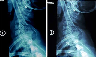
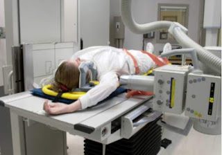
If the patient’s condition permits then the arm is raised on the side closest to the image receptor. The opposite shoulder is depressed so that the CR passes through the level of the coricoid process. If the patient has upper extremity injuries, then the opposite arm is raised and the shoulder closest to the film is depressed (reverse Swimmer’s
.
In this picture the patient is positioned for a reverse Swimmer’s view. Either arm may be raised depending on the patient’s condition
.
a full cervical spine lateral Swimmer’s view. The three contour lines can be drawn for assessment of alignment; however, the detail of the posterior architecture and apophyseal joints of C7/T1 are more difficult to distinguish. Some radiologists require the full spine Swimmer’s view because the vertebrae are more easily counted. Unless there is accuracy in identifying the structures and number of the vertebrae, the coned Swimmer’s view may not be helpful. In such cases the radiologist must be consulted since a CT scan may be necessary to completely evaluate the spine
.
.
The two radiographs above are of the Swimmer’s view in which the entire cervical spine is imaged. The alignment of the vertebrae and the apophyseal joints can be assessed, and the vertebrae are easily counted for accuracy
.
.
Truescene is your right choice

السبت، مايو 9

Cases 21-30


case21
.
A 43-year-old woman presents with red lesions on her hands. Her family history is significant for systemic lupus erythematosus affecting her mother. Physical examination reveals erythematous- to-violaceous papules on the dorsal surface of all fingers, including both the proximal and the distal interphalangeal joints. The papules are asymptomatic. Examination with a magnifying glass reveals telangiectasia involving the proximal nail folds. A facial rash is also noted. The patient denies any systemic complaints, including muscle weakness
.
.
Answer
.
This patient was diagnosed with Gottron’s papules, which are associated with dermatomyositis. Classified as a connective tissue disease, dermatomyositis presents with various cutaneous manifestations, including proximal nail fold telangiectasia, Gottron’s papules, and heliotrope rash (a purplish erythema and edema of the malar and periorbital regions of the face). Proximal muscle weakness may precede or follow the onset of skin findings. Systemic involvement
correlates with elevated muscle-associated enzymes. Treatment usually entails use of high-dose oral steroids and may also require antimalarials and immunosuppressive agents
.
.
case 22
.
An 82-year-old man presents for evaluation and treatment of a slowly enlarging growth on the right side of his neck. He states that the growth is not tender but occasionally drains a foul-smelling, cheesy substance. Examination of the site reveals a 3 cm by 3 cm, freely movable dermal nodule. On closer inspection, a central pinpoint opening on the skin surface is noted. Pressure on the lesion results in expression of a malodorous, caulk-like material
.
.
Answer
.
This patient has an epidermoid cyst, an invagination of the skin characterized by the accumulation of cellular debris and keratin within an epithelial-lined sac. Over time, the material develops a pungent odor. These lesions are most commonly found on the back and chest but can arise on virtually any part of the body. Treatment utilizing incision and drainage will effectively shrink the lesion, but recurrence is common unless the entire wall is excised. Epidermoid cysts have no malignant potential
.
.
case 23
.
A 45-year-old woman presents with a widespread, itchy, red rash on her trunk, neck, and extremities that began approximately two days ago. She denies any history of a similar condition, as well as any swelling of the lips or difficulty breathing. Her family physician recently prescribed a thiazide diuretic for hypertension and mild leg edema. Examination reveals a diffuse, erythematous, urticarial eruption involving more than 50% of her skin surface
.
.
Answer
.
This patient’s history and clinical presentation are classic for a drug hypersensitivity reaction. Antibiotics (most notably amoxicillin) and diuretics are common causative agents. A reaction to medication should be suspected in any patient who develops a morbilliform or urticarial skin rash. A detailed medical history and complete list of medications are of utmost importance in establishing causality. Immediate cessation of the offending agent usually results in prompt clearing. Oral antihistamines and topical steroids may hasten resolution. More severe cases may warrant a short course of oral prednisone
.
.
case 24
.
A 72-year-old woman developed a small, asymptomatic papule on her left arm several weeks ago. Since then, the papule has rapidly expanded in size. Today?s examination finds this 2-cm nodule with its raised, erythematous border and crusted center. Scattered actinic keratoses are noted on the patient?s hands and face. Axillary lymph nodes are not detected. Her past medical history is positive for basal cell carcinoma affecting her forehead
.
.
Answer
.
The lesion was removed by curettage, and biopsy confirmed a diagnosis of keratoacanthoma. This tumor, which is considered a variant of squamous cell carcinoma, is characterized by a rapid growth phase and the presence of a dome-shaped nodule with a central keratin plug or ulceration. Sun-exposed areas are most susceptible. Although many cases will spontaneously involute, there have been occasional reports of metastatic spread. For this reason, most clinicians recommend surgical removal or, when this is not feasible, radiation therapy
.
.
case 25
.
A 23-year-old man presents for evaluation of an asymptomatic growth under his right great toenail. He states that the lesion has been slowly increasing in size for several years and that he is now seeking treatment because of the cosmetic disfigurement. Physical examination reveals toenails that appear normal except for a ridge alongside the right great toenail. Noted is a shiny, pink, firm, subungual tumor protruding beneath the nail of this toe. Palpation does not elicit tenderness
.
.
Answer
.
Biopsy revealed that the lesion was a digital fibroma. These benign connective tissue growths usually present as a smooth, dome-shaped, solitary nodule, most often on the great toe. Multiple lesions may be associated with tuberous sclerosis. Antecedent trauma is sometimes correlated with onset. Digital fibromas can exert pressure on the nail matrix, resulting in distortion of the nail. Treatment, when requested for either cosmesis or relief of discomfort, entails surgical excision of the tumor
.
.
case 26
.
An 81-year-old, fair-skinned woman presents for evaluation of lesions that she has had for about two years on both legs. She admits to ample sun exposure in the past and has had a skin cancer removed from her face. Although the lesions are largely asymptomatic, several have bled following minor trauma. An examination of the patient?s legs reveals mild edema as well as multiple erythematous patches and thin plaques of varying sizes
.
.
Answer
.
Disseminated superficial actinic porokeratosis (DSAP) presents as multiple flat to barely elevated brownish-red patches on the extremities. The inheritance pattern of the disorder is autosomal dominant with variable penetrance. In genetically predisposed individuals, chronic sun exposure is implicated in the development of lesions, which tend to appear beginning in the fourth decade. Most cases can be diagnosed based on clinical appearance; histopathology will confirm the diagnosis. Uncommonly, DSAP may evolve into squamous cell carcinoma. Some lesions may respond to cryosurgery or topical application of 5-fluorouracil or imiquimod cream
.
.
case 27
.
An obese 76-year-old nursing home resident suffers from a recurrent blistering rash on her abdomen. The patient is immobile and frequently scratches the lesions. She is currently on oral medications to control hypertension and diabetes. On physical examination, tense bullae scattered on the abdomen are noted in addition to excoriations and superficial ulcerations
.
.
Answer
.
Punch biopsy acquired from a newly developed lesion revealed bullous pemphigoid. This chronic condition involves deposition of immunoglobulins within the dermis resulting in subepidermal tense blisters. The average age of onset is 65, with an equal incidence in males and females. This patient was treated with ultra-high potency topical steroids and secondary infection was prevented with topical mupirocin ointment. More extensive disease often requires systemic prednisone for adequate control
.
.
case 28
.
A 70-year-old man presents for evaluation of an asymptomatic scalp lesion that has been increasing in size over the past six weeks. His medical history includes actinic keratoses that have been treated cryosurgically with liquid nitrogen. Examination of the mid-scalp reveals a 1.8-cm indurated plaque with central scaling. Multiple keratoses are noted elsewhere on his scalp as well as on his face. Cervical lymph nodes are not palpable
.
.
Answer
.
Atypical fibroxanthoma most commonly presents as a reddened, dome-shaped nodule that arises rapidly on the head or neck of an elderly individual. Most cases appear to be related to chronic sun exposure or localized radiation therapy. Differential diagnosis includes squamous cell carcinoma, malignant melanoma, and angiosarcoma. Although the majority of lesions treated with simple curettage or shave excision do not recur, spread to lymph nodes has been reported, prompting some clinicians to recommend removal by either full excision or Mohs micrographic surgery
.
.
case 29
.
An 86-year-old resident of a nursing home has a chronic blistering rash on her hands and feet. She has multiple medical problems and is on several oral medications. Examination of her palms and soles reveals scattered flattened bullae and slightly indurated, well-demarcated, erythematous plaques. Several nails manifest dystrophy and hyperkeratosis, and coarse scales of her scalp are noted as well
.
.
Answer
.
The patient has acral pustular psoriasis. An uncommon variant of psoriasis, this chronic condition is characterized by brightly erythematous plaques and pustules, the latter sterile in nature. The nails are frequently affected. Adequate control may require administration of systemic agents such as acitretin, cyclosporine, and methotrexate. A combination of oral psoralen and ultraviolet light (PUVA) may also prove beneficial, as may the so-called "biologics" etanercept, efalizumab, and alefacept
.
.
case 30
.
A 66-year-old woman presents with an enlarging lesion of her right axilla, which she believes to be a wart. She reports that it bled recently, prompting her to seek medical attention. The patient is a cigarette smoker with no history of skin cancer. Examination of the affected area reveals a 3- x 5-cm erythematous plaque with a hyperkeratotic, crusted core. Cervical and axillary nodes are not palpable
.
.
Answer
.
 Cutaneous biopsy of this lesion revealed invasive squamous cell carcinoma. This second most prevalent form of skin cancer is rarely seen in sun-protected skin folds. Cigarette smoking is a risk factor. If left untreated, up to 20% of cases may metastasize. The uncommon location in this case illustrates the need for total body skin examination

Episode 4


Cervical spinal nerves
.
.
One of the most important understandings the technologist must gain is an appreciation for the fact that injury to a vertebra does not necessarily mean that there is an injury to the spinal cord; neither does injury to the spinal cord require a vertebral fracture for correlation. Neurological examination of the patient and radiographic interpretation by a radiologist is what determines what further testing is needed before a definitive diagnosis can be given. In many case instances the radiologist in consultation with a neurosurgeon may recommend a CT scan, and possibly magnetic resonance imaging, or even fluoroscopy guided spine motion studies. The point here is that the radiographer should be aware that injuries to spinal nerves or to “soft” tissue are issues in trauma care as well as awareness of bony injury. Traumatic injuries to the spinal cord or the spinal nerves may occur during a trauma event. 
.
.
A basic understanding of the gross anatomy of spinal nerves as part of ones understanding of the scope of trauma imaging and patient care is in order. In addition to the anatomy of the spinal nerves, the technologist should be aware that some sensory nerves carry more than just pain or the absence of pain; there is also touch, pressure distinction, two-point discrimination, temperature, proprioreception, and other sensory and motor functions. The complete picture of the patient is gathered by the physician performing the clinical evaluation, augmented with diagnostic testing. A brief review of the gross anatomy of the spinal nerves that exit the spinal cord is beneficial to ones imaging skills. It is difficult to know when an image is sufficient for diagnosis unless one knows what the elements of the diagnosis entail. Certainly a complete survey of the bony relationship to the spinal nerves and vertebral canal are necessary. Therefore, we should review the gross anatomy of the spinal nerves.
.
.
The gross anatomy of the spinal nerves is that there are thirty-one pairs of spinal nerves having attachments to the spinal cord. These are arbitrarily divided into 8 pair of cervical spinal nerves, 12 pair of thoracic nerves, 5 pair of lumbar nerves, 5 pairs of sacral nerves, and one coccygeal nerve. The eight pair of cervical nerves exit the spine in the following way: the first cervical nerve exits above the first cervical vertebra between it and the skull, the second cervical nerve exits below the first cervical vertebra between C1 and C2, the third cervical nerve below the second cervical vertebra, the fourth cervical nerve from below the third cervical vertebra between C2 and C3, the fifth cervical nerve from below the fourth cervical vertebra, the sixth cervical nerve from below the fifth cervical vertebra, the seventh cervical nerve from below the sixth cervical vertebra, and the eighth cervical nerve from below the seventh cervical vertebra between it and the first thoracic vertebra. Therefore, to effectively demonstrate potential injury to the eighth cervical spinal nerve the radiographer must demonstrate the vertebral body, apophyseal joints, and quadrilateral posterior architecture of both C7 and T1 vertebrae. The alignment of the 7th cervical and 1st thoracic vertebrae must also be demonstrated
.
.
the pattern for the exit of the eight pairs of cervical spinal nerves from the vertebral canal. These are mixed nerves carrying sensory and motor distributions
.
.
The spinal nerves exit the vertebral column through the intervertebral foramina. Each intervertebral foramen is formed by the pedicles of adjacent vertebrae, which have notches on their superior and inferior borders. The inferior vertebral notch and superior vertebral notch of adjacent vertebrae form the intervertebral foramen (arrows-picture below). Within each foramen lies a dorsal root ganglion of a spinal nerve
.
.
wait for the next episode soon on your TRUE SCENE

Episode 3

Soft Tissue Structures of the Neck
.
.
the soft tissue structures of the neck are just as important as the bony spine. This is especially true when we consider trauma imaging. The potential for soft tissue injury from penetrating trauma, blunt trauma, forces associated with motor vehicle accident, and other types of trauma heighten the risk for soft tissue injury. By soft tissue structures we imply that in addition to the bony spine, all tissues within the boundaries delimited by skin (anteriorly, posteriorly, and laterally). These tissues include muscles, fat, the airway, esophagus, blood vessels, and the like. For our study we will only consider the airway and the carotid and vertebral arteries
.
The airway is important to trauma because it must be maintained as part of the ABCs of survival (airway, breathing, and circulation). Radiographically, the injury to the airway or retropharyngeal space may indicate substantial trauma. Any deviation of the airway and other soft tissue structures may indicate a need for acute emergency treatment. It is necessary for the technologist to know some basic anatomy of the airway and identify its important features on a radiograph
.
.
Fascial planes described in the neck
.
Three fascial planes are described in the neck: investing, pretracheal, and prevertebral fascia. The investing fascia is a layer that encircles the structures of the neck. It is firmly attached to the mastoid processes, zygomatic arches, mandible, hyoid bone, and spinous processes, manubrium, clavicles, and both scapula. It is important because it contains the jugular venous arch and some muscles of the neck

Along the anterior portion of the neck is a thin layer of cervical fascia called the pretracheal fascia. It extends from the thyroid cartilage (C4) into the thorax. It is a split fascia which encloses the thyroid gland, trachea, and esophagus. Trauma to the trachea that results in a rupture of the trachea, or of a bronchus in the thorax can result in air seepage into the pretracheal fascial space. An acute swollen neck caused by air in the pretracheal fascia (mediastinal emphysema) may spread into the face along this route. 

The prevertebral fascia covers the muscles of the vertebral column, extending from the base of the skull to about the third thoracic vertebra. Its importance to us is that it is firmly attached to the anterior longitudinal ligament which can contribute to its injury from trauma. Because the pharynx is surrounded by a buccopharyngeal fascia there is a space created between it and the prevertebral fascia called the retropharyngeal space. This space contains the great vessels from the aortic arch, the heart, the trachea, the thymus and part of the esophagus. This space is important because it allows for movements of these structures, such as the esophagus during swallowing, and the beating of the heart. Trauma causing perforation into this space is radiographically significant; therefore, the radiographer must have an understanding of the dynamics of this anatomy so that it is included on routine plain films
.
.
The picture above demonstrates the relative locations of the fascia, particularly noted is the retropharyngeal space, which is a potential space. Blood or pus that infiltrates into this space can spread to the superior mediastinum
.
.
The airway
.

The airway begins just posterior to the nasal and oral cavities with the pharynx. The pharynx is the upper respiratory tract, but is common to both the digestive and respiratory systems. It conducts food to the stomach via the esophagus, and air to the lungs via the trachea. The airway can be seen in the picture above. Visualization of the airway is important to imaging of the traumatic cervical spine.

Vertebral and carotid arteries
.

There are four main arteries that pass through the neck in route to the brain to supply it with blood. These vessels are: the right and left vertebral arteries, and the right and left carotid arteries. An understanding of the gross anatomy of these vessels is important to radiographers because in certain types of trauma they could be injured. The goal of this brief review is to make the radiographer keener to the need of the patient toward other diagnostic test or surgery. It is not intended that the radiographer make a decision on patient care; however, valuable time should not be lost trying to get a good plain film radiograph when a CT scan may be quicker and more informative. An example would be to rule out a cervical spine injury from a fall, secondary to a gunshot injury. Now let’s consider the anatomy of these vessels.
.

The carotids and vertebral arteries in the neck have their origins from the great vessels arising from the aortic arch in the superior mediastinum. 
.
The blood supply to the brain from the neck is very important to survival because there are only four vessels that supply the entire brain with oxygenated nutrient rich blood. These vessels are the right and left vertebral arteries, and the right and left internal carotid arteries. Within the transverse foramina coarsens the right and left vertebral arteries in route to the brain supplying it with oxygenated blood. Each vertebral artery originates from its respective subclavian artery in the root of the neck (picture above). The vertebral arteries enter the transverse foramen of C-6 to ascend upward through the transverse foramina of C6-C1 entering the skull through the foramen magnum Through anastomoses these four vessels form contributories to the Circle of Willis supplying the brain with oxygenated blood. Any disruption in this supply can have grave consequences to the individual. Traumatic injury to these vessels can be evaluated by agiography, or in some cases with intravenous contrast CT angiography

.
.
.
The bony spine protects the vertebral arteries; but only muscles and soft tissues of the neck protect the carotid arteries as the picture below shows. Injury to blood vessels like the carotids and vertebral arteries must be considered in certain types of traumatic injury to the neck or chest

الجمعة، مايو 8

Episode 2

Atypical Cervical Vertebrae
.
.
Three of the cervical vertebrae are classified as atypical. These are the first cervical vertebra called the atlas, the second cervical vertebra called the axis, and the seventh cervical vertebra called the vertebra prominens
.
The Atlas
.
The first cervical vertebra is called the atlas because it articulates superiorly with the base of the skull, and bares its weigh. The atlas is atypical because it lacks pedicles, laminae, and spinous process. Instead it has an anterior and a posterior arch. Two large lateral masses lie between the arches having on their superior and inferior surfaces the four articular processes and their facets. The picture below demonstrates the two large superior articular processes, one on each lateral mass, which through their facets articulate with the occipital condyles on the skull base

The large lateral masses and their superior articular processes are seen. They are connected by an anterior and a posterior arch. The atlas does not have a vertebral body, pedicles, laminae, or a spinous process, which makes it atypical
The superior articular processes and occipital condyles form two joints called the atlantooccipital joints. These joints provide flexion and extension movements between the neck and skull such as in motioning “yes” by nodding of one's head

the upper cervical spine demonstrates the atlantooccipital joints formed by the condyles of the occipital bone and the superior articular processes of the atlas. The arrows point to the atlantooccipital joints which are superimposed on the lateral view

The Axis C2
.
The second cervical vertebra is also atypical in that the body of the vertebra is expanded bearing a tooth like process called the odontoid process (dens). It is the strongest of the cervical vertebrae. It possesses large superior articular processes for articulation with the atlas that rests upon them. Its parts are a bifid spinous process, inferior articular processes, superior articular processes, transverse processes and transverse foramina, pedicles, and a vertebral body that bares an expanded tooth-like process called the odontoid process
.
.

The superior articular processes of the axis, along with the two inferior articular processes of the atlas, form two atlantoaxial joints. A transverse ligament holds the odontoid peg of C2 in place against a facet on the posterior wall of the anterior arch of C1. These joints are functionally important because this is where the atlas and skull and rotate as a unit upon the axis. It is a side-to-side rotation as in disapproval nodding, which is contraindicated in acute head and neck trauma because such movement can cause spinal cord injury if certain types of traumatic fracture(s) involving the cervical vertebrae are present
.
.
The drawing (right) and lateral radiograph (left) demonstrates the odontoid process of C2 which articulates with the anterior tubercle and transverse ligament of the atlas
.
The atlantoaxial joints are also radiographically important, therefore, the radiographer should understand their relationship to normal anatomical function. These joints are aligned like all vertebrae along the lateral margins. Because these joints are diarthrodial joints, they are spaced between the articular processes by a synovial membrane and synovial fluid. Therefore, they should have a spacing that reflects this anatomy
.
Vertebra prominens C7
.
The seventh cervical vertebra is considered an atypical cervical vertebra because of its long spinous process that can be seen and felt in the back of the neck. Also, it has long transverse processes resembling a thoracic vertebra, but is distinguished by the absence of rib attachment. The vertebral arteries do not pass through the transverse foramen of the seventh cervical vertebra. Therefore the atypical vertebrae are C1, C2, and C7
.
.
wait for next episode on true scene by dr bassem el baz

Tension Headache


Tension Headache
.
.
Presentation
.
The patient complains of a dull, steady pain, described as an ache, pressure, throb, or constricting band, located anywhere from eyes to occiput, perhaps including the neck or shoulders. Most commonly, the headache develops near the end of the day, or after some particular stress. The pain may improve with rest, aspirin, acetaminophen, or other medications. The physical exam will be unremarkable except for cranial or posterior muscle spasm or tenderness
.
.
What to do
.
 * Perform a complete general history (including environmental factors and foods which precede the headaches) and physical examination (including a neurological examination).
  * If the patient complains of sudden onset of the "worst headache of my life," accompanied by any change in mental status, weakness, vomiting, seizures, stiff neck, or persistent neurologic abnormalities, suspect a cerebrovascular cause, especially a subarachnoid hemorrhage, intracranial hemorrhage, or arteriovenous malformation. The best initial diagnostic test for these is computed tomography, but when CT is not available and the patient does not have papilledema or other signs of increased intracranial pressure, rule out these problems with a lumbar puncture.
  * If the headache is accompanied by fever and stiff neck, or change in mental status, you need to rule out bacterial meningitis as soon as possible, again with lumbar puncture.
  * If the headache was preceded by ophthalmic or neurologic symptoms, now resolving, suggestive of a migraine headache, you may want to try sumatriptan or ergotamine therapy. If vasospastic symptoms persist into the headache phase, the etiology may still be a migraine, but it becomes more important to rule out other cerebrovascular causes.
  * If the headache follows prolonged reading, driving, or television watching, and decreased visual acuity is improved by viewing through a pinhole, the headache may be due to a defect in optical refraction, curable with new eyeglass lenses.
  * If the temples are tender, check for visual defects and myalgias that accompany temporal arteritis.
  * If there is a history of recent dental work or grinding of teeth, tenderness anterior to the tragus, or crepitus on motion of the jaw, suspect arthritis of the temperomandibular joint .
  * If there is fever, tenderness to percussion over the frontal or maxillary sinuses, purulent drainage visible in the nose, or facial pain exacerbated by lowering the head, consider sinusitis.
  * If pain radiates to the ear, be sure to inspect and palpate the teeth, which are a common site of referred pain.
  * Finally, after checking for all these other causes of headache, palpate the temporalis, occipitalis, and other muscles of the calvarium and neck, looking for areas of tenderness and spasm which usually accompany muscle tension headaches. Keep an eye out for especially tender trigger points which may resolve with gentle pressure or massage.
  * Prescribe anti-inflammatory analgesics (ibuprofen, naproxen), recommend rest, and have the patient try cool compresses and massage of any trigger points.
  * Explain the etiology and treatment of muscle spasm of the head and neck.
  * Volunteer the information that you see no evidence of other serious disease (if this is true); especially that a brain tumor is unlikely. (Often this is a fear which is never voiced.)
  * Arrange for followup. Instruct the patient to return to the ED or contact his own physician if symptoms change or worsen
.
.
What not to do
.
 * Do not discharge without followup instructions. Many serious illnesses begin with a minor cephalgia, and patients may postpone urgent; care in the belief that they have been definitively diagnosed on the first visit.
  * Do not miss subarachnoid hemorrhage and meningitis. (If you are not obtaining a majority of negative CTs and LPs, you may not be looking hard enough
.
Discussion

Headaches are common and most are benign, but any headache brought to medical attention deserves a thorough evaluation. Screening tests are of little value--a laborious history and physical examination are required. Other causes of headache include carbon monoxide exposure from wood heaters, fevers and viral myalgias, caffeine withdrawal, hypertension, glaucoma, tic douloureux (trigeminal neuralgia) and intolerance of foods containing nitrite, tyramine, xanthine. Tension headache is not a wastebasket diagnosis of exclusion but a specific diagnosis, confirmed by palpating tenderness in craniocervical muscles. ("Tension" refers to muscle spasm more than life stress.) Tension headache is often dignified with the diagnosis of " migraine" without any evidence of a vascular etiology, and is often treated with minor tranquilizers, which may or may not help. Focal tenderness over the greater occipital nerves (C2, 3) can be associated with an occipital neuralgia or occipital headache, and be secondary to cervical radiculopathy from cervical spondylosis. These tend to occur in older patients and should not be confused with tension headache. Remember to probe for the patient's hidden agenda. "Headache" may often be the justification for seeing a physician when some other physical, emotional, or social concern is actually the patient's major problem


EPISODE 1


                      Spinal Cord Injury

Spinal cord injury (SCI) is an injury to the spinal cord or nerve tracts that relay signals to and from the brain and body. Trauma is the leading cause of spinal cord injury and is caused most commonly by motor vehicle accidents. Falls and violent acts account for the second leading causes of spinal cord injury. Traumatic spine injury occurs in approximately 1 in 10,000 individuals per year; most are males in their teens or twenties. Greater than half of cord injuries occur in the cervical spine region, a third in the thoracic region, and the remainder in the lumbosacral area. Paramount to vertebrae injury is the question of neurological injury. Most cases of spine injury do not involve permanent cord injury. The standard of medical care demands a high index of suspicion of injury in certain type types of trauma. This means that in certain types of trauma the safest medical strategy is to assume an unstable fracture or dislocation of the vertebrae exists until proven otherwise. This point of view must be part of the working assumption of the entire medical team providing care to a trauma patient. As part of the medical team continuum of patient care, the radiographer must also assume spine injury until proven otherwise.

The management of a patient with a potential spinal cord injury begins in the pre-hospital environment under the care of paramedics. They are specially trained to identify common situations in which a spinal cord injury is likely to occur and correctly immobilize the patient. Ideally, the patient is immobilized in the neutral position on a full spine board with a combination of a cervical collar, side head supports, and strapping of the shoulders and pelvis so that the neck is not the center of the body's rotation. The spine is protected at all times during transport because it is not the center of the body's rotation should the patient move involuntarily. Sandbags and a cervical collar alone do not provide maximum protection when moving the patient, the shoulders and pelvis must be strapped to the board also. When the patient is moved to a gurney or x-ray table the spine must stay in alignment and not be rotated. The rule for protecting the spine from further injury is to immobilize it. These precautions are the standard of care for handling a trauma patient suspected of spine injury. 

Once the patient arrives in the hospital trauma suite, the patient may be removed from the spine board to a rigid transfer board. Logrolling the patient is the standard maneuver for any movement of the patient. This transfer process allows physical evaluation of the back (alignment of the spinous processes), rectum for bleeding, and other data. Under no circumstance should the log-roll technique be short changed by too few movers. The radiographer should not participate in the logroll procedure unless properly trained. If a transfer from the spine board is done it must be accomplished under the direct guidance of the trauma physician(s). The process can use nursing, emergency medical technicians (EMT), and providers from the trauma team. To properly logroll the patient there must be at least two persons on each side of the patient, and one at the head end to support the neck. The procedure should be smooth, and the entire spine kept in alignment during the movement.

To help understand the severity of spinal cord injuries and the radiographer's role in providing diagnostic images of the spine, a brief review of the structure of the vertebrae and spinal cord is essential. In this module we will explore current imaging standards and recommended imaging protocols for radiographic examination of the cervical spine and its spinal cord segments in critically injured patients. Although institutions vary in the type and functionality of radiographic equipment, trauma imaging standards can be completed using any available equipment. The key to a good radiography department’s response to trauma is the education of the technologists dealing with trauma. By remaining updated in current radiographic imaging standards the technologist is armed with the understanding of what it is to have a high suspicion for injury that translates into safe quality patient care


Anatomy of the Cervical Vertebrae
There are seven cervical vertebrae that are numbered from top to bottom (C1, C2, C3, C4, C5, C6, and C7). They are conveniently labeled as typical or atypical according to their anatomical features. The typical cervical vertebrae have the following parts: a vertebral body, two pedicles, two lamina, and a single spinous process, two transverse processes, two inferior, and two superior articular processes. Vertebrae three through six are typical cervical vertebrae. The first, second, and seventh cervical vertebrae are atypical. Regardless of whether a vertebra is typical or atypical there are some features shared by all cervical vertebrae that distinguish them from other vertebrae like the thoracic, lumbar, or sacral vertebrae.

Typical Cervical Vertebrae (C3-C6)

A typical vertebra is conventionally divided into two parts: a large anterior portion called the body and posterior to it the vertebral arch. The body bares the weight of the trunk, whilst the arch protects the spinal cord and its associated nerve roots. The vertebral arch is formed by two pedicles that extend from the posterolateral portion of the vertebral body joining the two laminae to make up the posterior bony ring. The posterior portions of the vertebral body along with its bony arch form a large foramen called the vertebral foramen. Successive stacking of the vertebral foramina forms a bony tube, called the vertebral canal, which encloses the spinal cord and its meninges. On each side of the vertebral arch, projecting laterally from the union of the pedicle and lamina is a transverse process. Extending posteriorly from the junction of the laminae is a single spinous process. The transverse processes and spinous process act as levers to which muscles attach to effect movement of the spine.

Also arising from the arch are four processes called zygapophyses that participate in forming joints of the spine. Two project superiorly and two inferiorly and are so named the superior and inferior articular processes. Each process bares a facet for articulation with an adjacent vertebra’s zygapophyses. The zygapophyses form joints called apophyseal joints. These articular surfaces are where joint movements of the spine occur

Articulations of superior articular processes of one vertebra and the inferior articular processes of an adjacent vertebra form a joint termed an apophyseal joint. Each articular process bares a facet, which is a smooth area on the bone where joint
articulation takes place. Each vertebra articulates with an adjacent vertebra above it and the vertebra below it to share four apophyseal joints, two above and two below. These joints are classified as diarthroses or synovial joints. Synovial joints provide free movement between the bones they join. They have an articular cartilage membrane surrounding a joint cavity. Within the membrane bound space is synovial fluid that reduces friction between the bones. In the spine these joints are capable of flexion, extension, lateral bending, and rotational movements
The posterior quadrilateral architecture and anterior vertebral architecture

Along with the apophyseal joints, the posterior quadrilateral architecture of each vertebra is anatomically important. This area encompasses those structures from the posterior boundary of the vertebral body to the spinous process. Its bony structures include the pedicles, laminae, superior articular processes, and inferior articular processes. The posterior quadrilateral architecture describes those structures that form the vertebral foramen as viewed on a lateral radiograph of the cervical spine.
The reason the posterior architecture is so important radiographically is that it involves structures that surround the spinal cord. A fracture involving the vertebral foramen could damage the spinal cord causing significant consequences for the patient. It is crucial to cervical spine imaging that the entire quadrilateral architecture is demonstrated for all cervical vertebrae and the first thoracic vertebra


The anterior architecture of the vertebra consists of the vertebral body for typical vertebrae and for atypical vertebrae C2 and C7. The boundary for the anterior architecture is the posterior and anterior contour lines. Structures to be found between these two lines are the vertebral bodies, intervertebral disc
, and ligaments that support the spine. Now we can look at the gross anatomy of the cervical spine as regions whose boundaries are formed by the anterior contour line, posterior contour line, and laminospinal line.

The intervertebral disc

The intervertebral discs are interposed between adjacent vertebral bodies placing them within the boundaries of the anterior architecture of the spine. The structure of an intervertebral disc consists of concentric outer rings of fibrous tissue called the annulus fibrosus. The annulus fibrosus provides the strongest attachment affixing vertebrae together. The center of the disc is a fluid moiety called the nucleus pulposus. It hydrostatically maintains the height of the vertebral column. The intervertebral disc is classified as a symphyses joint, which affords slight movement


The fluid moiety (nucleus pulposus) in the center of the intervertebral disc is seen. Surrounding the nucleus is a dark band that is the annulus fibrosus. Notice how the
annulus is tightly bound to the vertebral bodies holding them together, whilst the liquid moiety maintains the height of the space between the bodies. Also notice how the anterior and posterior longitudinal ligaments are firmly attached to the bodies and intervertebral discs
Anterior to the discs and vertebral bodies is a tightly bound anterior longitudinal ligament. It extends from the anterior surface of the sacrum to the anterior tubercle of the atlas and portions of the skull. Its role is to limit hyperextension of the vertebral column. The posterior longitudinal ligament is firmly fixed to the posterior periosteum of the vertebral bodies and intervertebral discs. It runs from the sacrum to the atlas and is within the vertebral canal. It functions to limit hyperflexion of the vertebral column
.

الأحد، مايو 3

CASES11-20


CASE11
  .    
A 94-year-old man presents complaining of acne. He has no major health problems and spends his free time fishing. Examination of his face reveals open comedones (blackheads) scattered on the nose, forehead, and cheeks. In addition, coarse facial wrinkles and furrows are observed. His medical history is negative for skin cancer and he denies experiencing any acne as a teenager
    
ANSWER
.
Favre-Racouchot syndrome is a condition characterized by the presence of open and closed comedones occurring within actinically-damaged skin. The disorder afflicts elderly individuals who have spent years in the sun. Many patients give a history of heavy cigarette smoking. The most common locations are the forehead and periorbital regions. Inflammatory lesions are not present. Extraction using a looped instrument (comedo extractor) provides cosmetic improvement. Prevention of new lesions is best accomplished with topical retinoids and sun protection
   .
CASE12
A 19-year-old African-American woman presents with an annular, reddened rash on her central forehead and upper nasal bridge. She states that the condition first appeared a few months ago as two discrete patches that later became confluent. The rash has remained asymp- tomatic. She denies any muscle or joint pain, recent sun exposure, or use of prescription medications. Physical examination finds an erythematous, arcuate plaque with slight induration and no scale. The surrounding skin appears normal
.
ANSWER
.
Erythema annulare centrifugum is classified as a gyrate erythema. It may occur in association with medications, systemic disease, infection, or malignancy, although many cases are never traced to a specific cause. Lesions begin as small erythematous papules that spread peripherally to produce polycyclic or annular plaques and can occur on the head, neck, trunk, or proximal extremities. Topical or intralesional steroids, or a combination of both, may hasten resolution of lesions. The differential diagnosis includes sarcoidosis, systemic lupus erythematosus, and erythema migrans
.
CASE13
A 65-year-old man complains of a painful lesion on his finger that appeared approximately three weeks ago. He gives a history of arthritis. Examination of the affected digit reveals a 0.5-cm translucent, shiny nodule with a slightly erythematous border situated on the distal phalangeal joint. Light palpation elicits tenderness and expresses a viscous fluid
.
ANSWER
.
A digital myxoid cyst is a flesh-colored-to-translucent papule or nodule located on either a finger or a toe and filled with hyaluronic acid, which has a jellylike consistency. Many cases arise secondary to friction or minor trauma and are associated with a history of osteoarthritis. Typical locations are the distal interphalangeal joint and the proximal nail fold. Asymptomatic cysts do not require therapy. Painful lesions may be drained, but they will frequently recur
.
CASE14
This two-year-old boy presents with an erythematous rash and bullae that developed within 24 hours. There was no antecedent inguinal or perianal dermatitis, but the rash was preceded by two days of low-grade fever, slightly decreased oral intake, and irritability. He takes no oral medications. Examination reveals mild inguinal lymphadenopathy, normal vital signs, and no evidence of dehydration. Mucous membranes are unaffected. Exquisite tenderness of the perineal skin is elicited. Nikolsky’s sign is positive on both bullae and surrounding skin. Ruptured bullae reveal moist skin with shallow erosions. A culture of aspirated fluid from intact bullae revealed no growth
.
ANSWER
.
This patient has localized staphylococcal scalded skin syndrome (SSSS). The condition is caused by toxigenic strains of Staphylococcus aureus and most commonly afflicts infants and young children. Its presention ranges from isolated bullae to generalized erythroderma. In the latter, toxin released during the inflammatory process leads to widespread desquamation of the epidermis. Toxic epidermal necrolysis (TEN) is included in the differential diagnosis. This is because both TEN and SSSS may exhibit a positive Nikolsky’s sign, in which gentle stroking of the skin leads to epidermal separation. However, TEN is often drug induced and uncommon in this age group. Localized SSSS responds to oral antibiotics such as dicloxacillin. More generalized cases require admission, intravenous antibiotics, and adequate hydration
.
CASE15
A 51-year-old man seeks consultation because his wife noted a new mole on his back. The lesion is asymptomatic. The patient gives negative family and personal histories for skin cancer and atypical nevi. He is in good general health and lives in a wooded area, where he spends much time outdoors and owns a variety of animals including dogs and horses
.
ANSWER
.
Ticks are blood-sucking arachnoids. Many different species exist; among the most commonly encountered by humans are the dog (Dermacentor variabilis) and deer (Ixodes scapularis) varieties. Ticks do not fly or jump, but crawl onto their hosts. Some species are capable of transmitting serious infections including Lyme disease, babesiosis, ehrlichiosis, and Rocky Mountain spotted fever. For this reason, ticks should be completely removed as soon as possible after discovery. This patient’s “mole” was eradicated with fine-tipped forceps
.
CASE16
A 16-year-old high school student developed a rash on his forehead six days ago. Two days after onset, he was seen by a family practitioner who prescribed cephalexin for a suspected staphylococcal infection. Despite treatment, the eruption continued to spread. He complains of mild fatigue but denies fever or swollen glands. He is a wrestler and has a match in two days. Examination of his forehead reveals multiple erythematous papules and papulovesicles
.
ANSWER
.
This patient is experiencing a primary episode of herpes simplex infection, probably acquired from skin-to-skin contact with another wrestler (herpes gladiatorum). He was advised of the contagious nature of the disease and that other wrestlers must not be exposed to active lesions. Herpes gladiatorum most commonly manifests as vesicular lesions on the head and neck. Primary infection may be accompanied by malaise, low-grade fever, and regional lymphadenopathy. This patient was treated with oral valacyclovir
.
CASE17
The parents of a one-year-old boy who has a rash on his back seek medical consultation. The eruption was first noted approximately four weeks ago, at which time a pediatrician recommended application of an over-the-counter 1% hydrocortisone cream twice a day. Subsequently, both of two affected areas began to enlarge. Examination reveals annular, erythematous patches with raised borders and central clearing. Family history is negative for eczema. Two kittens live in the house
.
ANSWER
.
Tinea corporis is characterized by well-demarcated, erythematous, scaling patches and plaques. Because a cell-mediated immune response results in central clearing, hyphae are best identified from the border. Many cases arise from direct contact with an infected dog or cat. The condition is often misdiagnosed as eczema and treated with topical steroids, which results in gradual extension of the lesions. Tinea corporis responds readily to treatment with an antifungal cream, although more extensive cases may benefit from an additional course of oral therapy
.
CASE18
A 14-year-old boy who says he has had a sore mouth for the past two days also complains of a slight fever, abdominal discomfort, and loss of appetite. His previous medical history is unremarkable. Examination of the oral cavity reveals erosive lesions on his tongue and palate. Also noted on his hands and feet are scattered, erythematous papulovesicles that are asymptomatic
.
ANSWER
.
Hand-foot-and-mouth disease (HFMD) is an infectious disorder characterized by vesicular lesions occurring within the mouth and on the hands and feet. Most cases are caused by strains of coxsackie virus. Epidemics among school-aged children are not uncommon. Oral lesions rapidly progress from macules to vesicles that become erosive and painful. Associated findings may include fever, malaise, abdominal pain, and anorexia. Signs and symptoms resolve within seven days. The differential diagnosis of HFMD includes aphthous ulcers and herpes simplex virus
.
CASE19
A 25-year-old African-American man presents with a chronic scalp condition. He has been treated with numerous oral antibiotics in the past without success. He complains of discomfort, pimple formation, drainage, and bleeding of the mid- and posterior scalp. Physical examination finds dusky erythema accompanied by follicular papules and pustules, as well as evidence of scarring alopecia. Some areas are tender to palpation and exhibit slight purulence
.
ANSWER
.
Folliculitis decalvans is a scarring alopecia associated with chronic infection of the scalp. Culture most commonly reveals growth of Staphylococcus aureus, although response to antibiotics is variable. The pronounced inflammatory infiltrate destroys the hair follicle, leading to scar formation and permanent hair loss within the involved areas. A 10-week course of therapy combining oral rifampin and oral clindamycin proved helpful in this case
.
CASE20
A 14-year-old girl complains of a discolored tongue. She denies sore throat, change in taste, or difficulty swallowing. Her medical history is unremarkable and she takes no oral medications. Physical examination finds whitened, irregularly shaped patches on the dorsal surface of her tongue. The papillae are not enlarged. The remainder of the oral cavity, including the upper and lower palate, exhibits no visible
abnormalities


ANSWER
.
Geographic tongue, or migratory glossitis, is a benign condition that presents with raised, white patches intermingled with reddened, atrophic areas on the dorsal surface of the tongue. The map-like appearance of the tongue gives the condition its name. Women are affected more than men and most patients are asymptomatic. A concomitant systemic or cutaneous abnormality is rarely detected, and there is no correlation with cigarette smoking. In most cases, the only treatment required is reassurance
.
WAIT FOR NEXT EPISODE ON TRUE SCENE BY DR BASSEM ELBAZ