Plain Film Radiographic Imaging of the Traumatic C-spine
Trauma imaging of the cervical spine begins with a high suspicion for vertebral or spinal cord injury. Standard radiographic views are obtained with the patient supine, fully supported on a spine board, with neck collar and immobilization apparatus in place. The standard trauma film series at most institutions are: a horizontal beam lateral and Swimmer's view (if necessary), anterior-posterior (AP) view, and open mouth odontoid view
.
Diagnostic imaging technologists should be aware of the indications for special views of the cervical spine such as: inability to adequately visualize all vertebrae (especially C2 and C7/T1), questionable fracture of articular pillar, a question of a fracture in the axial plane, a question of possible fracture fragments near the cord. Additional views if needed are the: Swimmer’s, Fuchs, and panorex views; and/or CT, and MRI
.
.
Besides knowing what the standard protocol is for trauma imaging of the cervical spine, assuring that the diagnostic criteria is met for each view taken is the overriding principle. This is important because the radiographer is usually the first to see the radiographic images and must make a decision on whether to submit the images for physician interpretation, or to repeat the imaging sequence until images with the diagnostic criteria is accomplished. Therefore, the advanced imaging specialist must know the diagnostic criteria for each view and assure that it is met in a timely manner. Also, the technologist must know when enough radiographs have been attempted and a consultation with the radiologist is appropriate since other imaging modalities may be needed to acquire sufficient diagnostic information. You should be aware that the radiographer is legally responsible for image quality and the radiologist for the acceptance of all radiographic images they interpret. Therefore, the radiographer must assure that they have met the diagnostic criterion for each view. Before the radiographer submits an image for diagnosis there are at least three things that must be well thought-out. The first is that the patient is properly positioned. Secondly, that the path of the central ray is correct. Third, has the diagnostic criteria for the view been met
.
.
.
Now what constitutes an acceptable lateral radiograph of the cervical spine? The radiographer should make sure that the field of view (FOV) includes from the base of the skull through the first thoracic vertebra. There should be minimal rotation and misalignment of the vertebrae. In addition, the radiographic technique should provide enough bony detail so that the three contour lines can be easily drawn. Soft tissue structures within the anterior and posterior margins of the skin should be included and not “burned out” on the film. Alignment of the three contour lines must be easily made from the film silhouette. The entire anterior and posterior architecture of the vertebrae must be must be visualized
Now what constitutes an acceptable lateral radiograph of the cervical spine? The radiographer should make sure that the field of view (FOV) includes from the base of the skull through the first thoracic vertebra. There should be minimal rotation and misalignment of the vertebrae. In addition, the radiographic technique should provide enough bony detail so that the three contour lines can be easily drawn. Soft tissue structures within the anterior and posterior margins of the skin should be included and not “burned out” on the film. Alignment of the three contour lines must be easily made from the film silhouette. The entire anterior and posterior architecture of the vertebrae must be must be visualized
.
Horizontal beam lateral Swimmer’s view
The Swimmer’s view is part of the routine imaging of the traumatic cervical spine whenever the distal cervical spine is not adequately visualized. 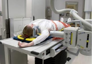 If the patient’s condition permits then the arm is raised on the side closest to the image receptor. The opposite shoulder is depressed so that the CR passes through the level of the coricoid process. If the patient has upper extremity injuries, then the opposite arm is raised and the shoulder closest to the film is depressed (reverse Swimmer’s
If the patient’s condition permits then the arm is raised on the side closest to the image receptor. The opposite shoulder is depressed so that the CR passes through the level of the coricoid process. If the patient has upper extremity injuries, then the opposite arm is raised and the shoulder closest to the film is depressed (reverse Swimmer’s.
In this picture the patient is positioned for a reverse Swimmer’s view. Either arm may be raised depending on the patient’s condition
.
a full cervical spine lateral Swimmer’s view. The three contour lines can be drawn for assessment of alignment; however, the detail of the posterior architecture and apophyseal joints of C7/T1 are more difficult to distinguish. Some radiologists require the full spine Swimmer’s view because the vertebrae are more easily counted. Unless there is accuracy in identifying the structures and number of the vertebrae, the coned Swimmer’s view may not be helpful. In such cases the radiologist must be consulted since a CT scan may be necessary to completely evaluate the spine
.
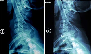 .
.The two radiographs above are of the Swimmer’s view in which the entire cervical spine is imaged. The alignment of the vertebrae and the apophyseal joints can be assessed, and the vertebrae are easily counted for accuracy
.
.
Truescene is your right choice
Truescene is your right choice


ليست هناك تعليقات:
إرسال تعليق